Anyone with a brain knows that our thoughts happen in our brains. But our metaphors belie our confidence in this assertion: “I know it in my heart,” “I have a gut feeling about it,” “I use my muscle memory to play this game.” Each of these statements says a little bit about what it feels like to process information. Do we know that our thoughts occur inside our heads because we can feel them happening there? Or do we feel them happening there because we’ve been taught very early on that our brains are where our thinking happens?
Egypt, India, China

We don’t really have any evidence of the opinions of prehistoric humans on where their seat of consciousness might reside. However, we can infer that they knew the brain was a vital organ, because it is evident from fracture patterns on prehistoric skulls that warriors tended to aim for the head if they wanted to kill their victims. The first written records on the functions of the various internal organs comes to us from the ancient Egyptians. They, like many other ancient societies, believed the heart to be the most important organ in the body, housing the soul and the conscience of the possessor. Embalmers charged with preparing a body for mummification removed and preserved the lungs, liver, stomach, and intestines and left the heart intact in the body, as it was needed in the afterlife for judgment to be passed on the deceased’s soul. The brain was removed and discarded.
For all their apparent brain disdain, the Egyptians were aware that head injuries could present symptoms in locations throughout the body. Contrast this with the physicians of ancient India, who associated a variety of illnesses with brain injury, all of which were localized to the head. The heart was again the most important of the organs in India, but Indian (Ayurvedic) medicine did place a special emphasis on cause-and-effect in medicine, as well as introducing a more scientific approach to medicine than was previously seen. Ancient India was also pioneering in its use of dissection to determine the functions of internal organs. However, much of their scientific observation was tied up with their metaphysics (a feature common to all civilizations of this time period).
Ancient China, with its ban on dissection, had an intricate physiology and anatomy, but one that was again based heavily on their metaphysical beliefs rather than direct observation. The system of Zang Fu proposed that the five most important organs (the Zang) were the heart, spleen, liver, lungs, and kidney, with the heart being the most important and the seat of consciousness. The Fu, on the other hand, were auxiliary organs. These included the intestines, bladder, gallbladder, stomach, and something known as sanjiao. The sanjiao was made up of the cranial cavity, thoracic cavity, and pelvic girdle, and was assumed to play a role in digestion and respiration. It’s clear that the brain wasn’t even on the radar of Chinese physicians as far as housing our thoughts is concerned.
Hippocrates, Aristotle, Galen
While many of the ancient Greeks believed that the conscious mind existed in the heart, Hippocrates (of Hippocratic oath fame) was one of the first to assert that the brain was the control center of the body. He and his followers noted that the optic nerve connected the eye directly with the brain, and several of his followers went on to associate the cerebellum with motor function and hypothesize that the complexity of wrinkles in the brain is proportional to one’s intelligence. Of course, dissections were still a rarity at this time, so many of these conclusions, accurate though they may be, are really pretty wild conjectures.
Aristotle, on the other hand, still contended that the mind resided in the heart. He reasoned that because the heart was beating, centrally located, and warm, that it must be “more alive” than the brain, which was cooler. He believed that the brain, with its extensive system of folds and blood vessels, acted as a sort of radiator to cool the blood from the heart. Since he knew that heat rises, it made sense to have a heat-dissipating organ at the top of the body.
The question would lay dormant for another few centuries until it was picked up again by Galen, a Roman physician in the 2nd century AD. He performed dissections and vivisections (dissections where the subject is still alive) on a number of different animals, closely examining their anatomy, especially of the nervous system. His work led him to believe that the brain controlled the body and that the nerves conveyed information back and forth from the body to the brain.
In his most striking demonstration, Galen performed a vivisection of a pig for an audience of physicians and anatomists, showing that when he cut the recurrent laryngeal nerves, the pig continued to struggle, but no longer squealed. All the doctors knew, either from their education or from their own work in anatomy, that nerves radiated outward from the brain to all other parts of the body. The squealing pig experiment was direct proof that those nerves must transmit information, in this instance from the brain to the larynx of the squealing pig. That meant that the brain itself must be telling the larynx to squeal. Though there were still brain-skeptics after this event, Galen’s dramatic demonstration was essentially the end of the belief that the heart controlled the body. It was also clear that the sequence of events for the squealing pig was: 1) The pig thinks “Squeal!” 2) The brain sends this thought via nerves to the larynx. 3) The larynx actually generates the squeal. The fact that the squeal originated in the pig’s brain led scientists and philosophers alike to assert that all thoughts must be generated in the brain, even those which don’t have a direct connection with motions of the body.
Where in the brain do thoughts occur?
While the structural anatomy of the brain advanced by leaps and bounds after Galen, the functions of various parts of the brain were subject to much speculation and little rigorous science until well into the Age of Enlightenment. One of the more notable of these speculators was Descartes, who believed that the mind and brain were separate entities. According to him, the mind was not a physical entity. It existed in some metaphysical realm, and communicated to the brain through the pineal gland. In his view, the brain was basically a middleman between the mind and the body. Another strange theory that arose during this period was phrenology. The basic tenet of this belief is sound: that different parts of the brain control different actions. However, phrenologists believed that those areas of the brain grew or shrank depending on whether someone was good or bad at a certain skill. Thus, phrenologists believed they could tell what you were good at, and even your personality, emotions, and mental afflictions by examining how lumpy your head was, and where the lumps were located.

By the early 1800s, phrenology had a number of adherents, but very little scientific support. Beginning in 1823, Jean Pierre Flourens began to perform what he called brain ablations, where he removed or damaged various parts of the brains of living animals (in this case, pigeons) and observed their behavior afterwards. He noted that removing the cerebrum caused loss of hearing, vision, and seemingly all sensation in the pigeons, whereas removing the cerebellum caused loss of motor control, and removing the medulla caused the pigeons to die. He also noted that if he removed large sections of the cerebrum, but not the whole cerebrum, the birds were still largely functional. He concluded from this that there exist three main regions of the brain (cerebrum, involved in sensation and thought; cerebellum, involved in motor control; and medulla, involved in vital functions like respiration and heartbeat) that operate in a connected way to one another.
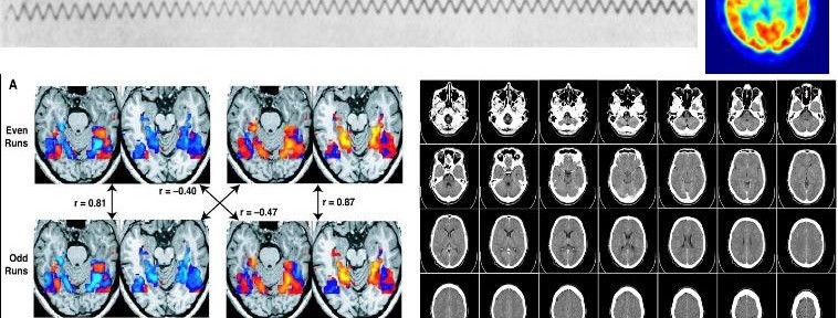
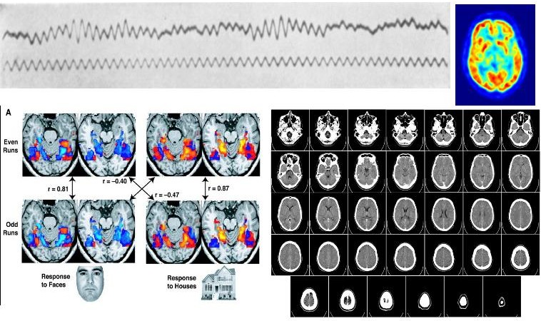
In 1861, Paul Broca presented evidence that patients suffering from aphasia (loss of ability to speak) all displayed brain lesions in the same area of the frontal lobe of the brain (now called Broca’s area, in honor of him). After this, studies associating damage of various regions to the brain with behavioral disorders began to pour in from scientists like Karl Wernicke and Alois Alzheimer. Other researchers, including Gustav Fritsch and Eduard Hitzig, used electrical stimulation of different parts of the brain to induce various responses, noting which parts of the brain controlled which actions. Around 1900, Oskar Vogt and Korbinian Brodmann began to use microscopy and newly developed ways of staining cells in order to show that the brain was made up of many different kinds of cells. In the 20th century, the types of localization studies described above proliferated and were refined by a multitude of scientists. The 20th century also saw the rise of techniques to probe the inner workings of the brain directly:
1) Electroencephalography (EEG) was developed by Hans Berger beginning in 1924.
2) Computed Tomography (CT or CAT scan) was developed over a period of 40 years and commercialized in the 1970s by Godfry Hounsfield.
3) Positron Emission Tomography (PET scan) was pioneered by James Robertson and colleagues in 1961.
4) Functional Magnetic Resonance Imaging (fMRI) was first performed on humans in 1992 to show changes in blood flow to various areas of the brain in real time.
Within the last 20 years, we’ve also seen the beginnings of working brain-machine interfaces in humans. Most of us would consider hooking our brains up directly to a computer to be a science fiction fantasy, but in reality, research in that direction is proceeding rapidly. Already, many hearing-impaired individuals are equipped with cochlear implants, devices that use electrical information from a microphone mounted on the outside of the head to directly stimulate the auditory nerve and produce the sensation of hearing. Experimental studies are ongoing with retinal implants to restore vision to severely visually-impaired persons, as well as with prosthetic limbs that can be directly controlled by the brain. Individuals that cannot physically communicate due to injury or disease have been able to have rudimentary conversations using EEG analysis.
These new developments promise to enable us to do amazing things in the future, while at the same time bringing up a lot of really tricky ethical questions. Those questions will need to be dealt with in time, but for now, human brain-machine interfaces are probably the clearest evidence we can get for an idea that Galen and Flourens first showed us with pigs and pigeons: that the brain is where thoughts occur.
References
- Chattopadhyāya, D. (1982) Case for a critical analysis of the Charak Saṃhitā In Studies in the History of Science in India (Ed. D. Chattopadhyāya). Vol. 1. New Delhi: Editorial Enterprises, pp. 209-236.
- “The Zang-Fu Theory.” Retrieved from http://www.tcmbasics.com/zangfu.htm
- Gadalla, Moustafa (2001). Egyptian Divinities – The All who are The One. Greensboro, N.C.: Tehuti Research Foundation, p. 78.
- S. Finger, Origins of Neuroscience: A History of Explorations into Brain Function, pp. 3-17.
- C. G. Gross. “Galen and the Squealing Pig.” The Neuroscientist. May 1998, Vol. 4, No. 3, pp. 216-221.
- J. M. S. Pearce. “Marie-Jean-Pierre Flourens (1794–1867) and Cortical Localization,” European Neurology. (2009) Vol. 61, No. 5, pp. 311-314.
- M. Loukas, et al. “Korbinian Brodmann (1868-1918) and His Contributions to Mapping the Cerebral Cortex,” Neurosurgery.(2011) Vol. 68. Issue 1. pp. 6-11.
- L. Haas. “Hans Berger (1873–1941), Richard Caton (1842–1926), and electroencephalography,” J Neurol Neurosurg Psychiatry. 2003 January; 74(1): 90.
- J. Clausen. “Man, Machine and in Between.” Nature. (2009) Vol 457. pp. 1080-1081.